TOR MAM

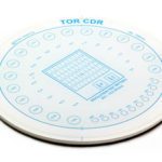
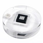
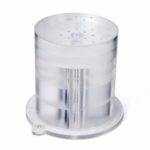
TOR MAM mammography phantom is designed to be used quickly and easily on a routine basis to provide an ongoing check of imaging performance, particularly those aspects which are most liable to deterioration. An ongoing record of these numbers will reveal any trend towards deterioration in imaging performance.

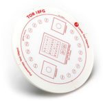
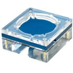
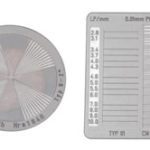
This test object is supplementary to TOR MAS or TOR MAX and provides a more “natural” image which may be preferred by radiographers and radiologists. The top (left) half contains a range of filaments, micro-particles and low-contrast details, representing pathological features in the breast. These are sensitive to the mammographic grey-scale, noise and unsharpness, and can be used to obtain an image-quality “score”. The lower (right) half simulates the appearance of breast tissue and contains micro-calcification in addition to fibrous and nodular details.

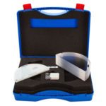
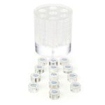
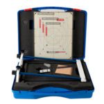
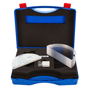
The kit consists of:
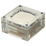
  • TOR MAM test object
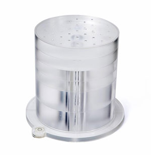
  • 7cm attenuator stack

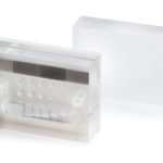
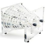
TOR MAM contains the following features:

  • 6 groups of multi-directional filaments.
  • 6 groups of micro-calcifications in ranges of 354-224, 283-180, 226-150, 177-106, 141-90, 106-93.
  • 6 groups of 3, low contrast details groups.
  • 6 groups of micro-calcifications (as in the above) with a clinically realistic breast tissue feature.
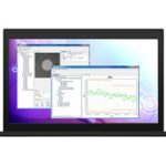
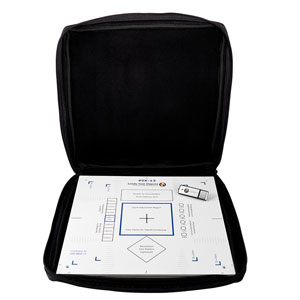
PIX 13

Leeds Test Objects’ PIX-13 phantom is designed to be used to check the image quality performance of digital radiography x-ray systems. These tests should be carried out at acceptance to establish a baseline, and on a routine basis, keeping an on-going record of the test results, which will indicate any deterioration in performance.

PIX-13 features the following test details:

  • Tube axis direction markings
  • Limiting spatial resolution (0.6 – 5.0 LP/mm)
  • Low contrast resolution details (6 x 15mm diameter details)
  • Dynamic range (7 different thickness Cu steps)
  • Unstructured are for placement of dosemeter
  • Unstructured area for homogeneity and signal adjustment (centre-marked)
  • Cassette size alignment markers (radiopaque)
  • X-ray to light field alignment markers (radiopaque)

PIX-13 should be used with either PMMA/Cu or Al attenuator plates (available separately).

The phantom is supplied in a soft protective carry bag.

Fluoro 4

Leeds Test Objects′ FLUORO-4 phantom is designed to be used to check the image quality performance of digital and conventional fluoroscopy x-ray systems. These tests should be carried out at acceptance to establish a baseline, and on a routine basis, keeping an on-going record of the test results, which will indicate any deterioration in performance.

FLUORO-4 features the following test details:

  • Tube axis direction markings
  • Limiting spatial resolution (0.6 − 5.0 LP/mm)
  • Low contrast resolution details (8 x 10mm diameter details, depths 0.4 − 4.0 mm)
  • Dynamic range (17 different thickness Cu steps each with a central 4.0mm diameter detail, depth 2.5 mm)
  • kV test-area (Yb 0.78mm)
  • Unstructured inner area (centre-marked)
  • Radiopaque numbers indicating dynamic range steps

FLUORO-4 should be used with either PMMA/Cu or Al attenuator plates (available separately).

The phantom is supplied in a soft protective carry bag.

Available in 2 Sizes:

FLUORO-4 (200x200mm)

FLUORO-4-L (300x300mm)


Conforms to standards:

  • DIN 6868-4
  • DIN 6868-150

Accu-Gold API

Accu-Gold-API_300x300The Accu-Gold digitizer and sensors are available with the “Accu-Gold” user software that enables simple measurement as well as rich visualization of the captured data.

For those applications requiring integration of Accu-Gold x-ray measurements with other automated equipment, Radcal provides an application programming interface (“API”).  The Accu-Gold API allows software developers to create their own applications.

The API is a Microsoft .Net 4 DLL compatible with most mainstream development languages for the PC platform such as Visual C++, C#, VB.Net, PowerShell, and LabView.  Contact Radcal for software licensing terms and conditions.

Skip to content