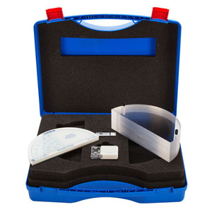
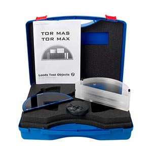
TOR MAS

A test object designed to be used quickly and easily on a routine basis to provide an ongoing check of imaging performance, particularly those aspects which are most liable to deterioration. An ongoing record of these numbers will reveal any trend towards deterioration in imaging performance.

The kit consists of:

  • TOR MAS test object
  • 7cm attenuator stack

Enables the following checks to be made:

  • Ten-step grey-scale plus two points for Sensitometric measurements.
  • Resolution limit (1.0 to 20.0 LP/mm) x 1
  • Low-contrast large-detail detectability (12 details, 5.6mm diameter)
  • High-contrast small-detail detectability (11 details, 0.5 and 0.25mm diameter)
  • Irregular-shaped particles on a step-wedge background (median sizes 125, 225, 325 microns representing micro-calcifications).
  • Low Contrast Resolution Pattern (1.8 to 5 line pairs/mm, representing filamentary structures).

TOR MAM

TOR MAM mammography phantom is designed to be used quickly and easily on a routine basis to provide an ongoing check of imaging performance, particularly those aspects which are most liable to deterioration. An ongoing record of these numbers will reveal any trend towards deterioration in imaging performance.

This test object is supplementary to TOR MAS or TOR MAX and provides a more “natural” image which may be preferred by radiographers and radiologists. The top (left) half contains a range of filaments, micro-particles and low-contrast details, representing pathological features in the breast. These are sensitive to the mammographic grey-scale, noise and unsharpness, and can be used to obtain an image-quality “score”. The lower (right) half simulates the appearance of breast tissue and contains micro-calcification in addition to fibrous and nodular details.

The kit consists of:

  • TOR MAM test object
  • 7cm attenuator stack

TOR MAM contains the following features:

  • 6 groups of multi-directional filaments.
  • 6 groups of micro-calcifications in ranges of 354-224, 283-180, 226-150, 177-106, 141-90, 106-93.
  • 6 groups of 3, low contrast details groups.
  • 6 groups of micro-calcifications (as in the above) with a clinically realistic breast tissue feature.