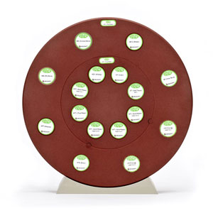
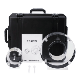
A computed tomography dose index (CTDI) QA phantom consisting of nested adult head and body segments.
TO CTDI permits the user to measure the dose index for computed tomography systems. The test object consists of two PMMA cylinders, one cylinder represents the head (160mm diameter) which, when nested inside the the other, represents the body (320mm diameter). The length of each cylinder is 140mm. Each cylinder contains 13.0mm diameter holes and comes with two adaptors tubes to permit the use of either 8mm, 10mm or 13mm diameter radiation detectors, used to take the dose measurements. There is a hole at the centre of the cylinders and one at each 90° interval, centred 10mm from the edge of the cylinder. TO CTDI includes nine PMMA pegs used to to fill the unoccupied holes and a phantom support for each size of cylinder.
TO CTDI has been designed in accordance with standards IEC 61223-3-5, IEC 61223-3-6 and IEC 60601-2-44.
TO CTDI enables the following tests to be made:
– CTDI100 (centre)
– CTDI100 (peripheral)
– CTDIw (weighted CTDI100)
– CTDIvol (weighted CTDIw)
– CTDIfree air
TO CTDI nested head and body phantom is supplied in a trolley case with a telescopic handle.
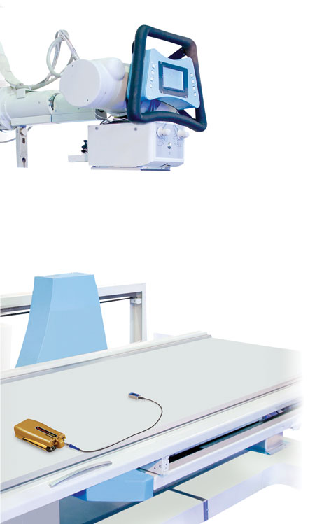
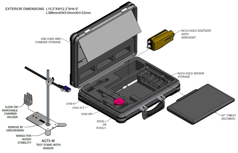
 Radcal is a premier provider of diagnostic radiation test instruments including the most comprehensive suite of x-ray sensors in the industry. Among these are an extensive selection of gold standard ion chambers including those tailored for CT quality assurance and maintenance. ACR and Joint Commission requirements demand that medical physicists measure beam width for today’s CT systems. Methods for performing this measurement either require special hardware and software or expensive consumables. Radcal is pleased to introduce The CTBW Tool Kit based on patent pending technology. This economical Toolkit works in conjunction with Radcal’s 10×6-3CT ion chamber to make beam width measurements with excellent precision. Two exposures determine a calibration factor and then beam widths for each additional collimation setting are calculated from a single additional exposure. Radcal’s free Accu-Gold Excel software automates the process and makes these measurements quick and easy.
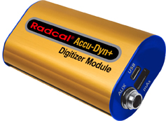 X-ray generators are manufactured to exacting standards that require precise measurement of the voltage (kV) and current (mA) applied to the x-ray tube. Radcal’s Dynalyzer product is the gold standard for measuring these quantities in the production environment. In the past, visualizing the data provided by the Dynalyzer required a digital display or scope. Radcal is pleased to announce the release of the Accu-Dyn+ solution which digitizes Dynalyzer results and makes them available in Microsoft Excel on a Windows-based tablet or personal computer. Analysis and reporting of kV behavior and practical peak voltage (PPV) has never been easier.
 To raise awareness of our profession, the International Organization for Medical Physics (IOMP) will celebrate this year, on November 7, the International Day of Medical Physics (IDMP).
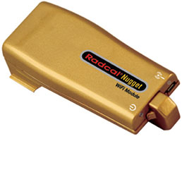
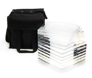
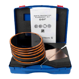 A set of phantoms for routine, commissioning and annual image quality QA for digital subtraction fluoroscopy systems.