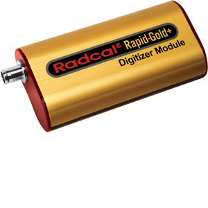
Rapid-Gold+
Highly Capable Solid-State Measurement System
The Rapid-Gold+ is the solid-state diagnostic x-ray measurement system in Radcal’s Accu-Gold family. Rapid-Gold+ systems encompass Radcal’s solid-state dose and multi-sensors along with optional current probes. Radcal’s unique stacked multi-sensors design provides unmatched accuracy and reliability along with reduced sensitivity to positioning errors. The Rapid-Gold+ is a balanced and cost-effective measurement solution.
The Rapid-Gold+ is a highly capable diagnostic measurement solution offering support for Radcal’s Solid-State sensors and advanced diagnostic functionality.
Systems Solutions
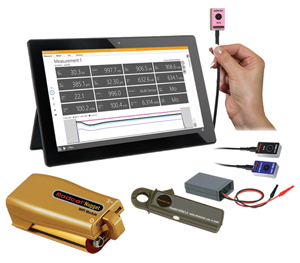
The Rapid-Gold+ provides a tailored solution to your individual diagnostic measurement needs when paired with:
• Radcal’s broad selection of Gold Standard sensors
• user friendly display that facilitates recording and reporting of results quickly and easily
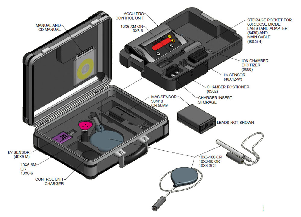
• carry case to transport your custom solution securely and conveniently
Sensor Selection
The Rapid-Gold+ supports Radcal’s full line of Solid-state Dose Diodes, and Solid-state Multisensors featuring the most accurate and compact stacked sensor design available.
Software and Display Technology
Radcal offers a number of display options including a compact tablet, a full-size tablet with keyboard, or you can use your own computer for a fully integrated and economical solution. In each case, the Accu-Gold and Accu-Gold Excel software provides a rich, user friendly, and automated environment in which to record, view, and archive your measurements.
Functionality
The Rapid-Gold+ provides a comprehensive set of parameters including Dose, Dose Rate, Waveform, Exposure time, kV, Filtration, HVL, and mA (optional).
Modalities
The Rapid-Gold+ is well suited for Radiography, Fluoroscopy, Mammography, CT, and Dental applications
 For use with general purpose electrometers, the 0.6cc thimble ionization chamber is ideal for dose measurements in modern wide beam multi-slice CT.

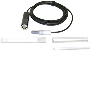
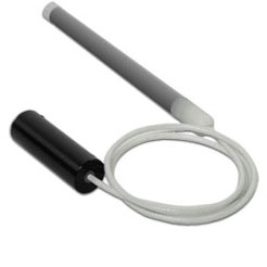 The RC3CT is an unsealed cylindrical, 10 cm pencil ion chamber with a 3 cm3 volume. It is designed specifically for CT x-ray beam measurements, either free-in-air or mounted in a head or body phantom. It has excellent energy and partial volume response along its entire 10 cm length.